Relationship between body weight and tissue thickness over the ischial tuberosity and the sacrum
Abstract
The thickness of tissue overlying the ischial tuberosity (IT) and the sacrum of 10 healthy young subjects were measured and compared with their body weight. The thickness of the tissue over the sacrum and the IT were found to be 13.8 mm and 43.4 mm. By grouping the body weight with increment of 5 kg, a linear relationship was observed between IT tissue thickness and body weight. However such relationship was not found for tissues over the sacrum. A better understanding on the tissue thickness over high risk bony area could help to design better pressure relief cushions.
Introduction
Pressure sores continue to be a major problem for wheelchair users and geriatric subjects. The common cause of pressure sore has been attributed to prolonged unrelieved pressure, which caused cessation of nutrient supply to the loaded tissues. Landis et al reported that interface pressure above 32 mmHg can cause local ischemia, and if prolonged will lead to ulceration1. The stresses experienced by the tissues are related to the thickness and properties of the tissue. Clark et al. reported that elderly with sores had less soft tissue over the sacrum. Garber et al3 found that thin patients have higher pressure over bony prominences than average weight and obese subjects. Brienza et al4 reported that higher sitting interface pressures were associated with a higher incidence of pressure ulcers in high risk elderly wheelchair users. In clinical situations, wastages of buttock tissues are commonly observed in chair bound individuals, and pressure relief cushions were usually prescribed to protect weight-bearing tissues against the onset of pressure ulcers. To achieve an optimal pressure distribution, it is important that the cushion material and surface contour should match the body shape and tissue properties of the user. By knowing the thickness and stiffness of tissues overlying bony prominence, and relating these information with the measured interface pressure magnitude, clinicians could have a better estimation on the stresses experience by the loaded tissues. Unfortunately, specialized equipment for tissue thickness measurement cannot be easily made available to seating clinics. An alternative means of assessing/predicting tissue depth is required.
RESEARCH QUESTION
The objective of this study was to document the depth of tissue overlying the ischial tuberosity and the sacrum were measured and compared to the body weight of the individuals. The findings of this work would provide information to the design of new generations of pressure-relief cushions.
Method
Ten healthy male subjects mean age of 22 years, ranged 21-30 years old, participated in this study. The depth of the soft tissues covering the ischial tuberosities and the sacrum were measured using a B-mode ultrasound system (Sonosite, USA). The measurements were conducted with the subject resting in a side lying position with hip and knee flexed at 90 degrees to simulate the sitting posture. Before the measurement, initial scan was applied to locate the bony prominence. Subsequently, five consecutive scans were conducted carefully to avoid any compression on tissues. The weights of the subject were measured using a clinical scale.
Result
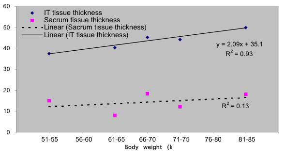 |
The weight of the subjects were 67 ± 9.6 kg. The average thickness of the left and right ischial tuberosities were found to be 43.0 ± 5.0 mm and 43.8 ± 5.4 mm respectively. The thickness of the tissue overlying the sacrum was 13.8 ± 5.0 mm. When the weight of the subjects were grouped and compared with the thickness of the tissue overlying the ischial tuberosities, a linear relationship was found (figure 1). However, such a relationship was not observed on the sacrum tissue.
Discussion
It is generally believed that by maintaining tissue bulk underneath bony prominence, the risk of pressure ulcers may be significantly reduced4. The thickness of tissues is thus an important factor to consider in pressure ulcer prevention. In clinical situation, measurement of tissue depth and stiffness were almost impossible due to the lack of suitable instrumentation. Without such information, assessment of the efficacy of tissue support materials can solely be based upon the magnitude and distribution of interface pressures, which does not really accounted for the properties of the loaded tissues. Recently, Apatsidis et al5 studied the effect of various materials on interface pressure at the seating interface of custom molded wheelchair seat and reported that viscoelastic foams produced lower peak-interface pressures and showed better pressure distribution than gels. However, clinical experience showed that viscoelastic foam does not always provide the best result to all wheelchair users. In fact, most of the related studies on cushion materials were conducted without accounting for the subject's tissue properties. In this preliminary study, it was demonstrated that body weight and depth of tissue at the ischial tuberosities are linearly related, but not for tissues at the sacrum area. This result provided clinicians with a simple way to predict the tissue thickness at the IT area by the subject's body weight. Further work is required to conduct measurement for chair-bound subjects, so that results could be generalized for application. The design of new-generations of pressure relief cushion should take into consideration of the tissue depth and tissue stiffness, so that these cushions can be optimized for better protection. Current work is undergoing to measure the stiffness of the tissues overlying bony prominence.
-
Ladis E.M., Micro-injection studies of capillary blood pressure in human skin. Heart, 1930; 15:209-228.
-
Clark M., Rowland L.B., Wood H.A., Crow R.A., Measurement of soft tissue thickness over the sacrum of elderly hospital patients using B-mode ultrasound, J. Biomed. Eng. 1989; 11(5):200-202.
-
Rondorf-Klym, L.M., Langemo, D., Relationship between body weight, body position, support surface, and tissue interface pressure at the sacrum, Decubitus 1993; 6(1):22-30.
- Garber S.L., Krouskop T.A., Body build and its relationship to pressure distribution in the seated wheelchair patient, Arch Phys Med Rehabil, 1982; 63:17-20.
- Brienza D.M., Karg P.E., Geyer M.J., Kelsey S., Trefler E., The relationship between pressure ulcer incidence and buttock-seat cushion interface pressure in at-risk elderly wheelchair users. Arch Phys Med Rehabil, 2001; 82:529-533.
-
Apatsidis D.P., Solomonidis S.E., Michael S.M., Pressure distribution at the seating interface of custom-molded wheelchair seats: effect of various materials. Arch Phys Med Rehabil, 2002; 83:1151-1156.
Eric Tam
Rehabilitation Engineering Centre
The Hong Kong Polytechnic University
Hunghom, Kowloon,
Hong Kong
852-2766-7670
852-2362-4365(fax)
rceric@polyu.edu.hk