Detect Soft-Tissue Stiffness Alteration in Denervated Human Tissue Using an Ultrasound Indentation Probe
Mohsen Makhsousa-d, Ph.D., Sam Perlmutterb,B.S., Ganapriya Venkatasubramaniana, M.S., Aditya Chawlaa, B.S., Yongping Zhenge, Ph.D., Fang Lina-c, D.Sc.
aSensory Motor Performance Program, Rehabilitation Institute of Chicago,
Depts of bPhysical Therapy & Human Movement Sciences, cPhysical Medicine & Rehabilitation, dOrthopaedic Surgery, Northwestern University, Chicago, IL, USA
eDepartment of Health Technology and Informatics, The Hong Kong Polytechnic University
ABSTRACT
Differences in soft tissue stiffness may provide a quantitative assessment and detection technique for pressure ulcers or deep tissue injury. To investigate this, a Tissue Ultrasound Palpation System (TUPS) was used to measure changes in soft tissue stiffness at the ischial tuberosity, greater trochanter, posterior mid thigh and biceps brachii from able-bodied persons and individuals with chronic spinal cord injury (SCI). Additionally, soft tissue thickness and soft tissue deformation during the manual indentation were measured at these locations. Significant differences in soft tissue stiffness were observed within the various anatomical locations tested in both groups. Differences in soft tissue stiffness were also observed between the able-bodied participants and the individuals with chronic SCI.
Key words:
soft tissue stiffness; deep tissue injury; ultrasound;
BACKGROUND
Current documentation and assessment of early pressure ulcer formation and severity are performed according to subjective impression from clinical inspection and palpation with appreciable inter-observer inconsistency. Much of the problem with this methodology stems from the frequency with which pressure ulcer formation begins well below the superficial layer of skin as a deep tissue injury (DTI). A DTI has been described by the NPUAP task force (1) as the development of significant pressure related damage under intact skin due to ischemia of the muscle bed. Full thickness punch biopsies have shown that lesions that could reasonably be described as a Stage I pressure ulcer in the NAPUAP and AHCPR staging systems demonstrate damage to deeper layers (2). These findings indicate that pressure ulcers sometimes start developing deep under the superficial layer of the skin. It was concluded that this kind of deep lesion can not be assessed using the current staging systems.
Soft tissue has been reported to have altered thickness and mechanical properties following a compressive load (3), post SCI (4), and in areas that are injured or adjacent to a pressure ulcer. One of the simplest interpretations of the mechanical properties is the stiffness of a material, i.e. the relationship between load applied to the material and the induced deformation. Therefore, it is possible that tissue stiffness be a clinically practical diagnostic tool to quantitatively detect, in an early stage, the pressure ulcer and possibly the DTI formation below intact skin. The purpose of this study was to investigate the feasibility of quantitatively measure tissue stiffness in individuals with SCI and compare with those who are able-bodied.
METHODOLOGY
Participants were twenty able-bodied subjects (Control group: 33.9±11.4 years, 68.8±9.4 kg, 169.6±6.6 cm, 23.9±2.8 BMI, 9 males and 11 females) and ten individuals with SCI (SCI group: 33.1±9.0 years, 76.7±16.4 kg, 173.8±7.0 cm, 25.4±5.1 BMI, 8 males and 2 females). For SCI group, averaged years post injury was 8.0±7.4 years and the level of injury spanned from C6 to T4.
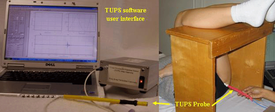 Figure 1. Experimental setup: The Tissue Ultrasound Palpation System (TUPS) was used to measure soft tissue stiffness in vivo and non-invasively in a simulated sitting position. The picture shows the testing of the IT on the right side of the subject. (Click image for larger view)
 A Tissue Ultrasound Palpation System(TUPS) (Figure 1) was used to test stiffness and thickness of soft tissue located at ischial tuberosity (IT), greater trochanter (GT), posterior mid thigh (MT) and biceps brachii (BIC). Data were collected bilaterally from IT’s and unilaterally from GT, MT and BIC. In the probe of TUPS, an ultrasound transducer measured the distance between probe tip and underlying bone surface. A 10 N load cell (ELFS-T3M, Entran Devices, Inc., Fairfield, NJ, USA) measured the force applied to testing site.
Participants were supine on examination table with lower legs on an elevated bench to maintain 90° flexion at both the hip and knee joints, representing joint configuration of a sitting posture (Figure 1). After applying the ultrasound gel to tip, the probe was placed on skin of targeted test location. Tissue was palpated with manually applied loading and unloading cycles to identify a clear echo or signal. Multiple echoes were visible due to reflection of sound waves from various layers of tissue (skin, fat, muscle, etc.). Only echo reflecting off bone was used to measure gross thickness of composite soft tissue layer. This was determined by identifying the echo with greatest amplitude and maximum displacement during cyclic loading/unloading. Each trial lasted 20 seconds and contained 5-7 loading cycles.
For each recording site, the unloaded thickness of soft tissue was obtained when loading force was zero. The soft tissue thickness (STT) was normalized to the corresponding BMI of each participant to minimize individual variance on body build. The maximal deformation of soft tissue was obtained at the trial time corresponded to the maximal loading force. The deformation was calculated as the percentage change relative to the initial unloaded thickness.
For each trial, recorded loading force and induced change of tissue thickness were used to establish a force-deformation relationship (Figure 2A). This relationship was used to estimate gross stiffness of composite soft tissue at each of the tested sites.
To estimate tissue stiffness from a non-linear force-deformation relationship (Figure 2A), we adopted a model of tissue elasticity composed of two parallel springs. Both springs originated from a common line with one spring being a distance of Xs shorter than the other (Figure 2B). Tissue stiffness was gauged in terms of three parameters, i.e. K1, K2, and Xs, where K1 and K2 denoted the stiffness of the two springs and Xs was the deformation of the spring K1 before loading of the spring K2. In the recorded data, this Xs corresponded to a tissue deformation before reaching the deflection point (Figure 2A). Mathematically, the relationship for these terms is described by following equations:
| F = K1X | x < xs | (Eq. 1) |
| F = K1x+K2(x-xs) | xs≤x≤xmax | (Eq. 2) |
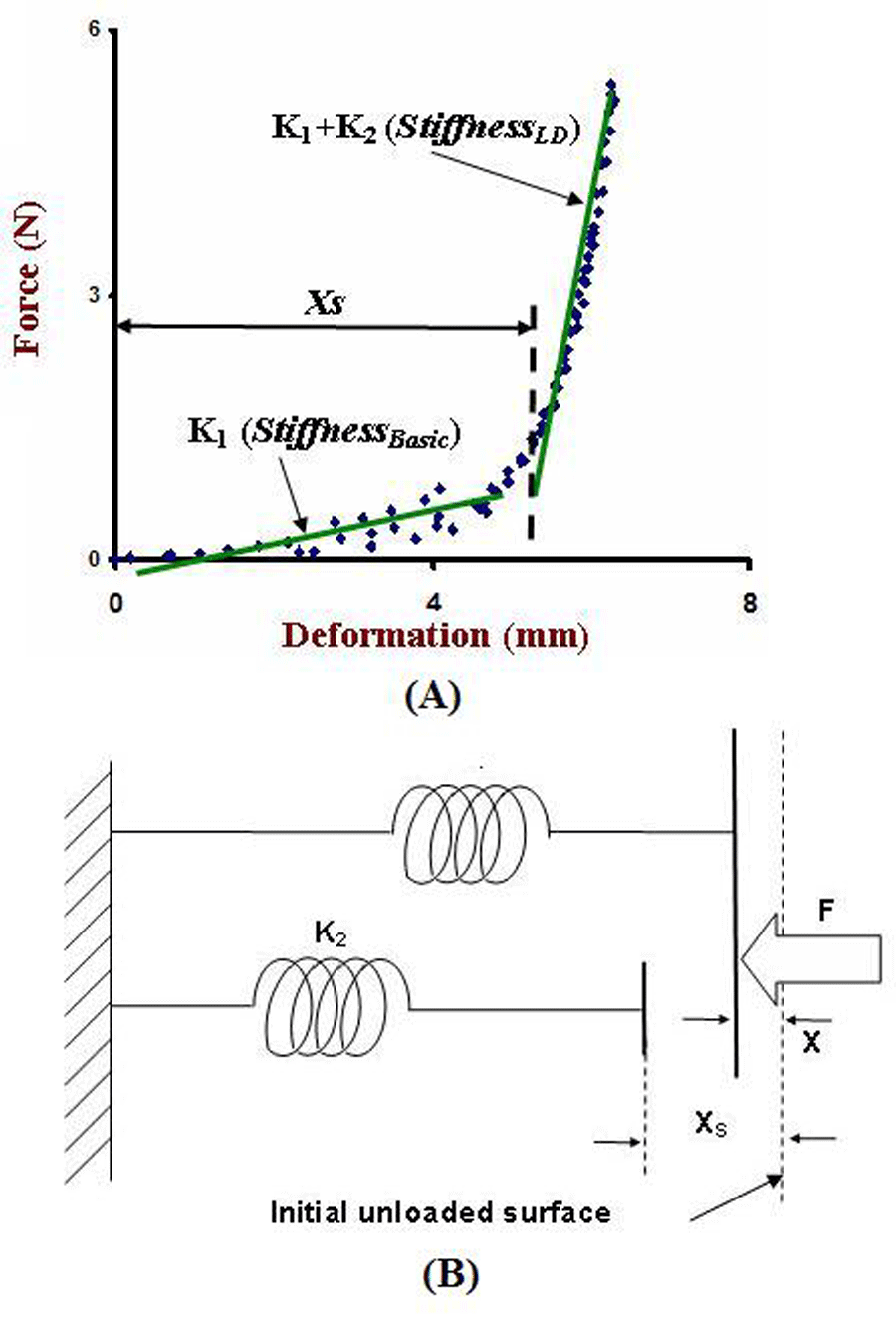 Figure 2: (A) A typical result of TUPS data recorded from GT from a subject. (B) Soft tissue material model (Click image for larger view)
 For each trial, the deflection point was determined for the force-deformation relationship and XS was obtained. Experimental data with deformation smaller than XS were fitted to Equation 1 to obtain K1. K1 and then defined as basic stiffness (StiffnessBasic), representing the slope of the first linear fit of the load-deformation curve (Figure 2A). Data larger than XS was fitted to Equation 2 to obtain K1+K2. K1+K2 was defined as the large-deformation stiffness (StiffnessLD), representing the slope of the second linear fit to the data. The large deformation was achieved by higher loading force. An example of this analytical approach is provided in Figure 2A.
For each location, mean and standard deviation were calculated for StiffnessBasic, StiffnessLD, Xs, STT (both absolute and normalized thickness), maximal loading force, and maximal deformation. A 2-sample t-test was performed for detecting significance of age, height, weight, and BMI between the Control and SCI groups. Paired t-test with 2 tails was used to determine significant difference of the data from the left and right IT. Two-way ANOVA was used to find overall significant effect of group (Control vs. SCI) and location (IT, GT, MT and BIC). When significance was found, a 2-sample t-test was used to obtain P-values for group effect on locations. When significance of location effect was detected, a paired t-test was used to obtain the P-values. To assess possible gender difference, an additional 2-sample t-test was used between all male and female participants. Statistical analysis was performed using SAS software (SAS® 9.3, SAS Institute, Gary, NC). Significance level was set to 0.05.
RESULTS
Subject Characteristics
There were no significant differences between the groups for age, height, or BMI.
Soft Tissue Thickness (Table 1)
Since no significant difference was found in tissue thickness collected from left and right IT (P>0.05), data from bilateral IT were averaged. In SCI group, BIC had the thickest soft tissue compared to other locations (P<0.001). In Control group, BIC was thickest, but only significant when compared to GT (P=0.020). GT was significantly the thinnest in both groups compared to BIC (SCI group: P<0.001; Control group: P=0.020) , as well as to MT in SCI group (P=0.005). BIC was significantly thicker in the SCI group compared to the Control group (P=0.040).
| Group | Able-bodied (N=20) | SCI (N=10) | ||||||
|---|---|---|---|---|---|---|---|---|
| Location | IT | GT | MT | BIC | IT | GT | MT | BIC |
| Thickness (mm) | 26.59±0.98 | 25.41±1.20 | 27.18±1.28 | 29.34±1.78 | 24.11±1.50 | 22.16±1.28 | 30.53±2.71 | 37.23±1.66 |
| P* | >0.05 | >0.05 | >0.05 | >0.05 | 0.006 | <0.001 | ||
| P^ | >0.05 | 0.020 | 0.005 | <0.001 | ||||
| P# | >0.05 | |||||||
| P | >0.05 | >0.05 | >0.05 | 0.004 | 0.040 | |||
| Normalized Thickness | 1.13±0.05 | 1.07±0.05 | 1.15±0.06 | 1.24±0.07 | 0.98±0.09 | 0.89±0.06 | 1.24±0.14 | 1.49±0.06 |
P* |
|
>0.05 |
>0.05 |
>0.05 |
|
>0.05 |
0.008 |
0.001 |
P^ |
|
|
>0.05 |
0.017 |
|
|
0.009 |
0.001 |
P# |
|
|
|
>0.05 |
|
|
|
|
P |
>0.05 |
0.024 |
>0.05 |
0.018 |
|
|
|
>0.05 |
P*: Significance for comparison with the value at IT. |
||||||||
Soft Tissue Deformation (Table 2)
Same as the tissue thickness data, deformation data from bilateral IT were averaged.
In SCI group, IT had significantly the largest deformation (GT P=0.011; MT P=0.006; BIC P=0.019). In control group, IT was significantly larger than GT and MT (GT P=0.014: MT P<0.001). Max compressive force of IT was significantly smaller among all locations in control group (GT P=0.003: MT P=0.004: BIC P<0.001). In SCI group, this was only significant when compared to BIC (BIC P=0.005).
| Group | Able-bodied (N=20) | SCI (N=10) | ||||||
|---|---|---|---|---|---|---|---|---|
| Location | IT | GT | MT | BIC | IT | GT | MT | BIC |
| Maximal Force (N) | 3.46±0.32 | 5.03±0.51 | 4.79±0.42 | 5.20±0.43 | 2.98±0.35 | 4.54±0.68 | 3.61±0.51 | 4.39±0.54 |
| P* | 0.003 | 0.004 | <0.001 | >0.05 | >0.05 | 0.005 | ||
| P^ | >0.05 | >0.05 | >0.05 | >0.05 | ||||
| P# | >0.05 | >0.05 | ||||||
| P | >0.05 | >0.05 | >0.05 | >0.05 | ||||
| Maximal Gross Deformation (%) | -37.48±2.55 | -31.98±2.71 | -25.84±1.37 | -34.07±2.73 | -33.96±3.04 | -26.64±2.44 | -23.25±2.31 | -27.08±1.64 |
| P* | 0.014 | >0.001 | >0.05 | 0.011 | 0.006 | 0.019 | ||
| P^ | 0.020 | >0.05 | >0.05 | >0.05 | ||||
| P# | 0.002 | >0.05 | ||||||
| P | >0.05 | >0.05 | >0.05 | 0.048 | ||||
P*: Significance for comparison with the value at IT. |
||||||||
Soft Tissue Stiffness (Table 3)
When comparing the 2 groups, StiffnessBasic at IT and MT were significantly lower for SCI group (IT: P=0.035; MT: P=0.001). StiffnessLD at BIC was significantly lower for SCI group (P=0.012) as compared to the Control group. Xs was significantly lower at GT (P=0.002) for SCI group compared to controls.
| Group |
Able-bodied (N=20) |
SCI (N=10) |
||||||
|---|---|---|---|---|---|---|---|---|
| Location |
IT |
GT |
MT |
BIC |
IT |
GT |
MT |
BIC |
| StiffnessBasicpo (N/mm) | 0.33±0.04 | 0.45±0.08 | 0.50±0.05 | 0.38±0.07 | 0.23±0.03 | 0.37±0.07 | 0.28±0.04 | 0.36±0.06 |
| P* | 0.037 | 0.003 | >0.05 | 0.043 | >0.05 | 0.026 | ||
| P^ | >0.05 | >0.05 | >0.05 | >0.05 | ||||
| P# | >0.05 | >0.05 | ||||||
| P | 0.035 | >0.05 | 0.001 | >0.05 | ||||
| StiffnessLD (N/mm) | 0.66±0.07 | 1.46±0.28 | 0.99±0.10 | 1.01±0.16 | 0.55±0.08 | 2.35±0.67 | 0.78±0.22 | 0.59±0.07 |
| P* | 0.007 | 0.004 | 0.018 | 0.013 | >0.05 | >0.05 | ||
| P^ | >0.05 | >0.05 | 0.007 | 0.012 | ||||
| P# | >0.05 | >0.05 | >0.05 | |||||
| P | >0.05 | >0.05 | 0.012 | |||||
| Xs (mm) | 4.79±0.47 | 4.74±0.53 | 3.85±0.32 | 5.17±0.44 | 4.68±0.37 | 2.84±0.28 | 3.74±0.41 | 4.87±0.34 |
| P* | >0.05 | >0.05 | >0.05 | <0.001 | >0.05 | >0.05 | ||
| P^ | >0.05 | >0.05 | >0.05 | <0.001 | ||||
| P# | >0.05 | 0.024 | 0.047 | |||||
| P | >0.05 | 0.002 | >0.05 | |||||
DISCUSSION
Investigation in this study was specifically aimed at finding potential differences in tissue stiffness and thickness, using the evaluated TUPS device, as a result of SCI and tissue denervation. Based on our data, tissue thickness was not significantly different in individuals with SCI from that of able-bodied persons in the areas of IT, MT, and GT. However, we did find that our participants with SCI had significantly thicker tissue at BIC. Upon eliminating the possible gender difference (no gender difference was found for tissue thickness in all participants), we think that the thicker tissue of BIC seen in SCI group may come from more use of their arms for propelling their wheelchairs.
Participants with SCI were found to have softer tissue in their buttock (IT) and thigh area (MT). The significantly lower stiffness may be attributed to several factors. Firstly, by having a chronic SCI, the muscular tissue around the IT is denervated and atrophied. Additionally, these participants may possess more fatty tissue in the buttock region from decreased levels of physical activity. Since a softer tissue may indicate an impaired capacity to resist compressive load, this finding may suggest a clue towards the vulnerability of tissue breakdown at the buttock/thigh region for those with SCI and sitting for a prolonged time.
The model used in this study was adapted from a model with similar parallel structure, which has been previously used for testing plantar soft tissue stiffness from individual with diabetes (5), and has been proven to be effective.
There are limitations in our study. The force applied to load the tissue is not constant across the different trials because the probe is operated manually. In addition, the indentation rates are also based on how fast the manual indentation is performed. However, it has been shown that the results are relatively insensitive to the indentation rates.
This study presents a novel data processing methodology for the TUPS. The outputs of the TUPS have been previously validated for different applications using a linear model (6). However, since our test involved applied large deformation of the soft tissues, the soft tissue force-deformation relationship obtained in our study showed non-linear behavior. Therefore, a non-linear material model for the stiffness data for our study was successfully developed and implemented.
REFERENCES
- Ankrom M, Bennet R, Sprigle S, et al. Pressure-related deep tissue injury under intact skin and the current pressure ulcer staging systems. Adv Skin Wound Care 2005;18(1):35-42.
- Witkowski J, Parish L. Histopathology of the decubitus ulcer. J Am Acad Dermatol 1982;6(6):1014-1021.
- Gefen A, Gefen N, Linder-Ganz E, et al. In vivo muscle stiffening under bone compression promotes deep pressure sores. J Biomech Eng 2005;127(3):512-524.
- Dupont-Versteegden E, Houle J, Gurley C, et al. Early changes in muscle fiber size and gene expression in response to spinal cord transection and excercise. Am J Physiol 1998;275:C1124-1133.
- Klaesner J, Hastings M, Zou D, et al. Plantar tissue stiffness in patients with diabetes mellitus and peripheral neuropathy. Arch Phys Med Rehabil 2002;83(12):1796-1801.
- Wendelken M, Markowitz L, Patel M, et al. Objective, noninvasive, wound assessment using B-mode ultrasonography. Wounds 2003;15(11):351-360.
ACKNOWLEDGEMENT
The project was supported in part by Medical Technology Systems and Falk Medical Research Trust.
Correspondence Author:
Mohsen Makhsous
Departments of Physical Therapy & Human Movement Sciences
Feinburg School
of Medicine
Northwestern Univ.
645 N. Michigan Ave., Suite 1100
Chicago,
IL 60611.
Phone 312-503-0073.
Email: m-makhsous2@northwestern.edu
Highlights
- Source Ordered
- No Tables
- Very Compatible
Gargoyles
Disney produced a television show in the mid 1990s called Gargoyles. It's a great show and I'm a big fan. A few years ago Disney started to release the show on DVD. The last release was of season 2, volume 1. That was two years ago. Volume 2 has not been released. Why? Poor sales. So if you should find yourself wanting to support my work, instead I ask you pick up a copy of season 2, volume 1. It's a great show and you might find yourself enjoying it.