Greg J. Williams1, Ashleigh Gonzales2, Alexander Lo1, Jamie Nolan1, Judy Grimmer1, Andrew Marquis1, Han Duerstock1, Susan Mendrysa1, Bradley S. Duerstock1
1Purdue University, 2Arizona State UniversityINTRODUCTION
Approximately 20% of the working population in the United States has a disability (Dept. of Labor, 2013). Yet only 2.7% of the science, technology, engineering, and mathematics (STEM) workforce report having a disability (Miner, Nieman, Swanson, Woods, 2001). Additionally, less than 3% of all biological sciences doctorates are earned by persons with disabilities (PWDs) (Supalo, Mallouk, Amorosi, Lanouette, Wohlers & McEnnis, 2009; NSF, 2013). There are several factors in the educational experiences of PWDs which help explain their underrepresentation in STEM professions: lack of independent hands-on experiences, low expectations, lack of role models, and limited exposure to science in and out of the classroom (Dunn, Rabren, Taylor, & Dotson, 2012; Supalo, Mallouk, Amorosi, Lanouette, Wohlers & McEnnis, 2009). Active practical STEM learning experiences in the science classroom or research settings are crucial to the development and success of scientists (National Research Council, 1996, Stefanich, 2007). Indeed, when undergraduates with visual and mobility disabilities were interviewed, the deterrent to them pursuing a laboratory-based career was not a lack of interest but the lack and inaccessibility of hands-on learning experiences (Duerstock, 2006; Mansoor et al., 2010).
In order to provide practical research experiences for PWDs including persons with blind or low vision (BLV) disabilities, it is necessary that each of the procedural components of the research task be accessible. Due to the foreseeable needs of a BLV graduate student entering into a biology research field, we decided to look at the accessibility of two common molecular biology procedures: micropipetting and transferring cultured cells from a well plate to a microscope slide.
In a survey of Purdue University life science research faculty, 85% of respondents stated that micropipetting was performed daily in their labs. Micropipetting is a ubiquitous component of most molecular biology protocols, including cell culturing. Over half of survey respondents performed cell culturing at least 2 to 3 times a week. In this study, test experiments were conducted to demonstrate the proficiency of BLV researchers micropipetting and culturing cells on a microscope slide coverslip in a well plate and then transferring it to a slide. Micropipetting liquids into and out of the wells required positioning the micropipette tip in the approximate middle of the wells near the bottom without touching the coverslip. Though this procedure is commonly performed by researchers with normal vision and hand eye coordination, it is extremely difficult to do while blind. Additionally, transferring cultured cells from the microscope coverslip in the well plate to a slide is normally accomplished using forceps without damaging the cells. This requires fine dexterity and good eyesight. For our blind researchers, both micropipetting and removing the coverslip from the well plate with forceps was impossible, even disregarding aseptic techniques.
A micropipette guide placed on top of the six well plate was designed to direct the tip to the proper location and depth. In addition, the micropipette guide would protect against contamination of the wells containing cell cultures. In order to remove the coverslip from the bottom of the well, a holder was designed that fit in the bottom of the well plate with a protruding handle for easy removal and transfer. 3-D printing facilitated a rapid and inexpensive iterative design process of building, assessing, and redesigning prototype solutions.
PURPOSE
The development of assistive technologies and methods promotes greater independence in the laboratory. The purpose of this research was to demonstrate how 3-D printing can be used for the rapid development of effective design aids to assist BLV researchers to perform common lab techniques, such as micropipetting and transferring cultured cells from a well plate to a microscope slide, requiring as little human assistance as possible. We assessed how well blind researchers were able to carry out these tasks compared to their sighted counterparts.
METHODS
Adaptive Aid Design Process
The well plate cover and coverslip holder were developed through a collaborative and iterative process using the SketchUp1 3-D modeling software for the design and a MakerBot® Replicator 2X2 for the 3-D printing. The design team consisted of the 3-D designers and developers, BLV end-users for testing, and experienced lab users of micropipetting and cell culturing lab techniques. Design iterations were examined by everyone and recommendations were made for printing another iteration.
Micropipetting
The first accessibility hurdle to overcome in micropipetting is the inaccessibility of the standard micropipettes themselves. The volume is set by turning a dial to the correct amount, but there are no reliable cues for a BLV person to set the volume. Alternatively there are fixed volume micropipettes, and combinations of these with braille labels for the volume were used to allow a BLV person to pipette the correct volume independently.
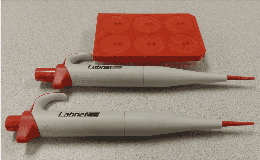 Figure 1: 3-D printed micropipette guide for six well plate (at top) and to fixed volume micropipettes (bottom) assist BLV users in dispensing liquids with less tactile probing
Dispensing and aspirating from a well plate without touching the bottom of the plate with the micropipette tip to avoid damaging the growing cells is possible for someone who is BLV, but it requires extensive touching of the well plate and micropipette tip. This is unacceptable in procedures which require aseptic technique. The solution was a well plate guide which fits on a standard six-well plate like the actual cover to prevent contamination. As shown in Figure 1, the guide has a recessed circle corresponding to each of the wells. Each well has two raised ports for dispensing and aspirating. The raised edge of the ports allows them to be easily found tactilely and for the tip of the pipette to be placed in them with minimal touching. This further guarded against accidental contamination.
The centered port is for dispensing. The hole is tapered to prevent the pipette tip from touching the bottom of the plate and damaging the cells. The off-centered port is for aspirating. The hole is angled to allow the pipette tip to get as near as possible to the bottom of the plate without touching it and to position it in the center of the coverslip where liquid is likely to collect.
Coverslip Holder
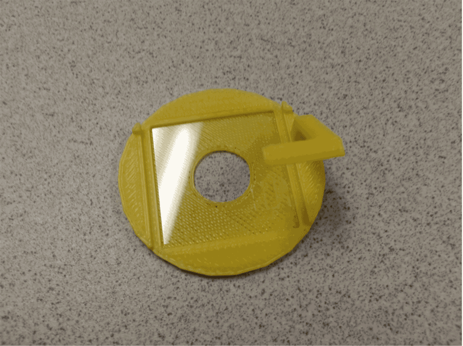 Figure 2: 3-D printed glass microscope coverslip holder with indentation to hold the coverslip in place. The diameter of the holder is equal to the diameter of the wells of the six-well plate.
Figure 2 shows the coverslip holder. The coverslip sits in the square indentation on the bottom of the holder. The handle is designed to allow easy removal of the coverslip from the well and is level with the top of the well plate so that the well plate cover and guide fit on securely. It is angled to keep it from interfering with the pipette tips during dispensing and aspirating using the well plate guide.
The grooves are to allow a glass slide to be laid on top of the coverslip. The two raised bumps at the edge of the holder are for easy orientation of the slide.
Experimental Evaluation
Once the tools were developed, we tested their liquid handling accuracy and potential for contaminating the wells during a series of mock cell culturing experiments. We conducted the procedures using a small cohort of blind and sighted researchers. The first experiment was designed to determine if there was a difference between the rate of contamination for blind and sighted researchers using the well plate guide. Two blind researchers and two sighted researchers, novices in micropipetting, pipetted 60 μL volumes of growth media into and out of a well plate using the guide. The plate contained two wells with coverslip holders. A control plate with deliberate contamination was also made. All plates were allowed to incubate over several days then inspected for contamination.
The second experiment compared the micropipetting accuracy of blind and sighted researchers using the well plate guide and coverslip holder. Two blind and two sighted researchers added and removed 65 μL of liquid from all six wells of a well plate with the guide and coverslip holder. As a control, a sighted researcher carried out the procedure without the guide and coverslip holder.
RESULTS
Table 1 shows the results of the first experiment. There was no statistical significant difference in the number of contaminated wells between blind and sighted researchers (ANOVA, P = 0.38). The most contamination was found in wells containing coverslip holders. Although they were wiped with alcohol and exposed to UV light for approximately 10 minutes, it is likely that they were the source of contamination and not the researchers.
|
Subjects: |
Well 1 |
Well 2 |
Well 3 |
Well 4 |
Well 5 w/ holder |
Well 6 w/ holder |
Blind 1 |
- |
- |
- |
- |
- |
+ |
Blind 2 |
- |
- |
- |
- |
- |
+ |
Sighted 1 |
- |
- |
- |
+ |
+ |
+ |
Sighted 2 |
- |
- |
- |
- |
+ |
- |
Control |
+ |
+ |
+ |
N/A |
N/A |
N/A |
The results of the second experiment showed that blind users had more errors of aspirating no liquids using the well plate guide. This is due to the inability of blind users to visually confirm that liquid was being withdrawn from the wells. Sighted users could double-check that liquid was being withdrawn. If these errors are discounted, there was no significant difference between all four researchers in liquid handling accuracy (one-way ANOVA, P = 0.065). However, using the guide resulted in significantly less liquid than not using the guide (control) (P < 0.0001).
DISCUSSION
Experiment 1 shows that blind researchers are as capable of carrying out sterile techniques as their sighted counterparts. It also indicates that the well plate guide does not introduce an additional source of contamination and aids blind researchers in maintaining a sterile environment. However, the coverslip holders have likely contributed to increased contamination. We do not know if the source of contamination is due to additional handling resulting from using the coverslip holders, inherent microscopic properties of the material that binds microbes, or inadequate sterilization prior to their use. More investigation is needed.
Experiment 2 focused on determining whether the well plate guide and coverslip holders assisted, hindered, or had no effect on proper dispensing and aspirating of liquids. The control results were much better than the results using the guide, even for sighted researchers. Modifications to the guide, such as changing the angle of the aspirating hole, may assist with this issue since blind and sighted researchers were equally effective if errors were ignored. Errors can be alleviated by more practice and possible verification techniques that liquid was aspirated.
Although the tools developed need improvement in micropipetting accuracy and sterile test performance, they were useful in allowing blind researchers for the first time to insert coverslips and add and remove liquids independently. A few simple adjustments should make the tools practical for a blind researcher to use in a cell culturing procedure. For example, the well plate guide can be adjusted for better tactile guidance of the pipette. The coverslip holders can be made with a more suitable material to prevent contamination. These are all straightforward changes that can be easily made when using a 3-D printer.
CONCLUSIONS
We believe better lab technique, which comes with practice would yield much better results and is likely a more important factor than modifying the design of the adaptive aids at this point. This study shows that inexpensive, yet precise tools developed through 3-D printing along with a clear understanding of the lab technique can enable BLV researchers to perform a cell culturing protocol independently and with confidence.
REFERENCES
Duerstock, B.S. (2006). Accessible Microscopy Workstation for Students and Scientists with Mobility Impairments. Assistive Technology, 18, 34-45.
Dunn, C., Rabren, K.S., Taylor, S.L., & Dotson, C.K. (2012). Assisting Students with High-Incidence Disabilities to Pursue Careers in Science, Technology, Engineering and Mathematics. Intervention in School and Clinic, 48, 47-54.
Mansoor, A., Ahmed, W., Samarapungavan, A., Cirillo, J., Schwarte, D., Robinson, J.P., & Duerstock, B.S. (2010) AccessScope Project: Accessible Light Microscope for Users with Upper Limb Mobility or Visual Impairments. Disability and Rehabilitation: Assistive Technology, 5(2), 143-152.
Miner, D., Nieman, R., Swanson, A.B., & Woods, M. (2001). Teaching Chemistry to Students with Disabilities: A Manual for High Schools, Colleges, and Graduate Programs (4th ed.). Washington, DC: The American Chemical Society.
National Research Council. (1996). In, National Science Education Standards. Washington, DC: National Academy Press.
National Science Foundation, National Center for Science and Engineering Statistics. 2013. Women, Minorities, and Persons with Disabilities in Science and Engineering: 2013. Special Report NSF 13-304. Arlington, VA. Available at http://www.nsf.gov/statistics/wmpd/.
Stefanich, G.P. (Ed.). (2007). Ontogeny of Inclusive Science. Dubuque, IA: Kendall-Hunt.
Supalo, C.A., Mallouk, T.E., Amorosi, C., Wohlers, H.D., & McEnnis, K. (2009). Using Adaptive Tools and Techniques to Teach a Class of Students Who are Blind or Low Vision. Journal of chemical Education, 86, 587-591.
United States Department of Labor, Bureau Of Labor Statistics, ‘Historical disability employment data,’ http://www.dol.gov/odep/topics/DisabilityEmploymentStatistics.htm, Web accessed on November 18, 2013.
FOOTNOTES
1SketchUp 2013 by Trimble Navigation Ltd. http://www.sketchup.com/
2MakerBot Replicator 2X by MakerBot. Phone: 347-334-6800. http://www.makerbot.com/
ACKNOWLEDGEMENTS
This project was supported by the National Institute of Health Director's ARRA Pathfinder Award to Promote Diversity in the Scientific Workplace (1DP4GM096842-01 to B.S.D.). Financial assistance was also provided by the State of Indiana through the Center for Paralysis Research at Purdue University. We wish to thank Debra Baluch from Arizona State University for assistance with the experimental test procedure.
Audio Version PDF Version