1Georgia Institute of Technology, Rehabilitation Engineering and Applied Research Laboratory (Atlanta)
ABSTRACT
This project tested the between-rater and within-session repeatability and reliability of the measurements from the MyotonPro device in a horizontal configuration on thin tissue over bony anatomy. The MyotonPro was chosen as an objective and instrumented alternative to manual palpation to monitor tissue properties for a number of clinical and research topics in rehabilitation. Stiffness, frequency, and damping data were obtained from 17 able-bodied subjects by two operators over the ischial tuberosity of the pelvis. The mean and coefficient of variation for stiffness are 175.10 ± 32.40 N/m, for frequency are 12.64 ± 1.15 Hz, and for damping are 0.790 ± 0.188 log, respectively. The between-rater reliability was excellent (ICC = 0.77 – 0.86). The within-session reliability of a single rater at the same location was similarly excellent for both operators (ICC = 0.94 – 0.97). Overall, these results demonstrate the MyotonPro device is capable of reliably taking measurements on thin adipose tissue over bony anatomy in a horizontal configuration in the hands of a novice user. For future studies, this tool could be used to classify buttock compliance as an indicator of general tissue health and may be useful in the assessment of risk and prevention of pressure ulcer formation.
INTRODUCTION
Prolonged durations of sitting are detrimental to the human body. Sitting causes compression of the tissue in the buttocks, which can lead to tissue damage. In cases such as spinal cord injury (SCI), individuals may not have the ability to stand, walk, or even perform weight shift maneuvers. The inability to remove the compressive forces on the tissues of the buttocks, back, and thighs puts this population at an increased risk of tissue damage and pressure ulcer formation. Typically, a skilled physician or clinician can manually palpate the buttock tissue to determine the tissue characteristics and susceptibility to damage. However, this hands-on approach requires extensive experience to detect subtle changes in the overall tissue characteristics between patients. The introduction of an objective assessment tool capable of quantifying mechanical properties would promote tissue monitoring of wheelchair users or otherwise sedentary individuals. The proposed tool would reduce the need for experienced palpations and minimize the subjective differences between examiners.
Many non-invasive devices exist to measure mechanical properties of soft tissue, including table-top test rigs [1,2] and handheld devices such as the Myotonometer [3-5] or algometer variants. [6] Tabletop testing rigs are highly accurate and precise [1,2], but too unwieldy to be used in a less controlled clinical setting. The majority of handheld devices rely on the application of a large force to determine stiffness by force-displacement [3-6] which may cause discomfort and further deterioration in cases of tissue damage. The MyotonPro device (Myoton AS, Estonia) was chosen for its size, measureable parameters, commercial availability, and general ease of use. It has been validated in other contexts, specifically using the device in a vertical configuration testing over muscle bellies to describe the state of tension of the underlying muscle. [7-9] For studying buttock tissue, the device needs to be held horizontally to measure the bulk tissue over the ischial tuberosity, as opposed to muscle belly tissue, with the participant in a side-lying posture to accommodate subjects with SCI. Therefore, the purpose of our work was to examine the intra- and inter-rater reliability of mechanical property measurements of bulk soft tissue over the ischial tuberosity. Specifically, the three questions we aimed to answer were: How repeatable are the within-session measurements by the same operator at the same location? Furthermore, how repeatable are the measurements across operators? Finally, how sensitive to probe placement are the measurements?
METHODS
A convenience sample of 17 able-bodied subjects were used for this study with IRB approval. Participants were asked to assume a side-lying position with hips and knees at 90° of flexion. The peak of the ischial tuberosity of the pelvis was palpated and marked using a skin marker. Four additional marks were added approximately 15-20mm superiorly, inferiorly, laterally, and medially to the center mark. The MyotonPro device was used to measure stiffness, frequency, and damping at each of the five marked locations, starting at the central point. A second measurement was then taken at the central point. After a minute of rest, a third measurement was taken at the central point, totaling seven measurements. The entire procedure was then repeated by a second operator.
The MyotonPro device delivers a series of five low-impact impulses of sub-Newton force to the skin via its 3mm-diameter probe. The acceleration data of the probe is measured and used to calculate the damped oscillatory response of the underlying tissue, approximated as a spring-mass-damper system. The device then outputs the oscillation curves, average stiffness (N/m), frequency (Hz), and damping (logarithmic decrement) values of the tissue from the five impulse-responses. Stiffness values represent the resistance to contraction or deformation, frequency values represent the intrinsic tension of underlying tissue, and damping values represent the ability of the tissue to recover initial shape after the impulse deformation. [10] The MyotonPro device is programmed to automatically flag any data sets with coefficients of variation (CoVs) over 3.0%. These high-CoV trials were excluded and re-measured immediately.
The collected data were downloaded from the device to proprietary MyotonPro software then analyzed in Minitab 18 Statistical Software (Minitab, Inc.). Descriptive statistics were calculated for each operator using all seven testing locations from each subject, summarized by means and standard deviations (Table 1). Intra-class correlation coefficients (ICCs) were calculated using an Excel spreadsheet available online [11] to quantify reliability of measurements within-session measurements of a single operator and between two operators. The data from the repeated tests on the central location, totaling three measurements per subject, were separated by operator. Intra-rater reliability ICC values were found for each operator by using the repeated test data for each subject in the Excel spreadsheet. Inter-rater reliability values were calculated by using the entire repeated central location data set for Operator 1 versus the same data set for Operator 2. As per Fleiss (2007), ICC values greater than 0.75 were indicative of excellent reliability, 0.40 to 0.75 as good to fair reliability, and less than 0.4 as poor reliability. [12] Finally, the sensitivity to probe placement was analyzed by computing CoVs of the parameters measured at the first central point and the four adjacent locations; the second and third tests at the central point were excluded from this analysis. The mean and standard deviation values for this data set were calculated (Table 1).
RESULTS
The convenience sample of 17 subjects (11 female) had a mean (SD) age of 25.94 (8.10) and BMI of 24.68 (3.80). Overall stiffness of the tissue above the ischial tuberosity ranged from 114.00 N/m to 277.00 N/m with a mean (SD) of 175.10 (32.40) N/m across all trials and participants. Frequency was 12.64 (1.15) Hz, and damping was 0.790 (0.188) logarithmic decrement.
Intra-rater Reliability
Within-session reliability of each operator was excellent for both operators (ICC = 0.94 – 0.97 for all reported parameters).
Inter-rater reliability
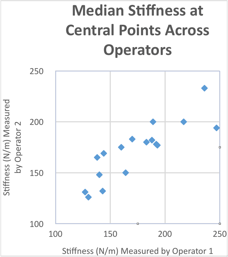
Tissue stiffness at the central location was highly correlated across operators (Figure 1, Pearson correlation value = 0.854). Furthermore, inter-rater reliability was excellent for all parameters (ICC = 0.77 – 0.86).
Placement Sensitivity
The CoVs for the data set containing the first central location and the four adjacent locations for each subject had values ranging from 8.36% to 27.45% (Table 1). Frequency was the most consistent. Damping was the least consistent and had the highest variability.
| Parameter | Operator | Mean (Standard Deviation) | CoVs (All Locations) | Intra-Rater ICC | Inter-Rater ICC |
|---|---|---|---|---|---|
| Stiffness (N/m) | 1 | 176.97 (35.91) | 20.11 | 0.95 | 0.86 |
| 2 | 173.26 (28.60) | 16.47 | 0.95 | ||
| Frequency (Hz) | 1 | 12.913 (1.199) | 9.41 | 0.95 | 0.77 |
| 2 | 12.366 (1.034) | 8.50 | 0.94 | ||
| Damping (Log) | 1 | 0.7481 (0.1239) | 16.41 | 0.95 | 0.81 |
| 2 | 0.8317 (0.2283) | 27.54 | 0.97 |
DISCUSSION
The MyotonPro device performed well in this horizontal configuration over non-muscle tissues. Within-session, each rater had excellent repeatability at the central points over the ischial tuberosity. Similarly, the reliability was excellent between raters. This reinforces the literature claiming high repeatability and reliability of the device. [7-9] The subject population had high variation as seen by the high CoVs of the stiffness and damping values. Variability was expected across the 15-20mm separation between adjacent testing locations. Naturally, proper probe placement is still integral to obtaining proper measurements.
It is important to note that time effects or trends were not considered in this study. The operators consistently tested in the same order without randomization. Operator 2 always tested after Operator 1 finished collecting data. Future work may involve a randomized operator order and testing over multiple visits to further investigate any potential time effects on the measurements.
CONCLUSIONS
The MyotonPro exhibited excellent repeatability and reliability in the hands of two novice operators, as reflected by the excellent intra-class correlation coefficient values. Measurements demonstrated sensitivity to probe placement for the 15-20mm separations between testing locations. All items considered, the device is adequately reliable for measurements in this previously-untested configuration over non-muscle tissue. These findings support the future use of the MyotonPro device to investigate the properties of buttock tissue.
REFERENCES
[1] Horikawa M. (2001). Effect of visual display terminal height on the trapezius muscle hardness: Quantitative evaluation by a newly developed muscle hardness meter. Applied Ergonomics, 32:473-478.
[2] Murayama, M., Nosaka, K., Yoneda, T., & Minamitani, K. (2000). Changes in hardness of the human elbow flexor muscles after eccentric exercise. European Journal of Applied Physiology and Occupational Physiology, 82, 361-367.
[3] Gubler-Hanna, C., Laskin, J., Marx, B.J., Leonard, C.T. (2007). Construct validity of myotonometric measurements of muscle compliance as a measure of strength. Physiol. Meas., 28, 913-924.
[4] Kerins, C.M., et al. (2013). Reliability of the myotonometer for assessment of posterior shoulder tightness. International Journal of Sports Physical Therapy, 8(3), 248-255.
[5] Jarocka, E., et al. (2012). Muscle stiffness at different force levels measured with two myotonometric devices. Physiol. Meas., 33, 65-78.
[6] Morozumi K, et al. (2010). A new tissue hardness meter and algometer; a new meter incorporating the functions of a tissue hardness meter and an algometer. J. Phys. Ther. Sci., 22, 239-245.
[7] Bailey, L., et al. (2013). Parameters Representing Muscle Tone, Elasticity and Stiffness of Biceps Brachii in Healthy Older Males: Symmetry and Within-Session Reliability Using the MyotonPRO. J Neurol Disord 1(1).
[8] Mooney, K., et al. (2013). Symmetry and within-session reliability of mechanical properties of biceps brachii muscles in healthy young adult males using the MyotonPRO device. Working Papers in the Health Sciences 1(3).
[9] Schneider, S., et al. (2015). Feasibility of monitoring muscle health in microgravity environments using Myoton technology. Med Biol Eng Comput 53(1): 57-66.
[10] MyotonPRO Digital Palpation USER MANUAL, Myoton AS, Tallinn, Estonia, 2016.
[11] Hopkins W. Analysis of reliability with a spreadsheet. A New View of Statistics, Internet Society for Sport Science. Available online: http://sportsci.org/resource/stats/xrely.xls; 2010.
[12] Fleiss JL. Reliability of Measurement. In: Fleiss JL. The Design and Analysis of Clinical Experiments. 1st ed, New York: Wiley and Sons; 2011.
[13] Makhsous, M., et al. (2008). Investigation of Soft-Tissue Stiffness Alteration in Denervated Human Tissue Using an Ultrasound Indentation System. J. Spinal Cord Med., 31, 88-96.
Acknowledgements
The authors would like to formally recognize and thank Dr. Deborah Wendland for her assistance with our study. Also, many thanks to Dr. Stephen Sprigle for providing us with the knowledge and experience vital to our data analysis.