Aquatin Hydrostander: A Novel Device for Pediatric Aquatic Physical Therapy
Kevin Ling, Brenda Burke, Jonathan Ptak, Juan Ayala, Daniela Valero, Mark Maynes, Leena Pramanik, Cooper Rickerson, Eric Fuller, Walter Murfee, Sarah Rowlinson
J. Crayton Pruitt Family Department of Biomedical Engineering, University of Florida
Problem Statement:
Aquatic physical therapy is a proven rehabilitation treatment for patients that suffer from peripheral limb movement disorders or other movement disabilities [1]–[6]. It is particularly helpful in providing therapy for children who suffer from an inability to maintain a healthy standing posture or control leg movements. Therapy in an aquatic environment lessens the stress of the patient's weight on their legs, enabling them to more easily control limbs and work on muscle strength [2], [4], [7]–[11]. However, current aquatic therapy methods for children or patients with mental disabilities often necessitate the presence of two physical therapists, one of whom must focus on holding the patient safely in place while the other engages the patient with the actual exercises. In analogous therapies that take place outside of a pool, standing frame devices are used to secure the patients in place, rather than a second physical therapist. Standing frames have been proven to be effective for allowing patients with lower limb disabilities to engage in proper muscle exercise [12]–[22]. The goal of this project was to design an analogous device that would function as a standing frame in an aquatic setting for use in aquatic physical therapy.
Solution:
We devised a special device that would serve the same function as a physical therapy standing frame but could be instead used in the aquatic pool setting. By taking advantage of the human body's natural buoyancy in the water, it is possible to maintain the correct therapeutic posture in the water with our device without requiring major input from the physical therapist. Thus, the physical therapist is freed to engage with the patient in other ways without worrying about holding the patient in one place.
We designated several functional requirements:
The device must be able to secure the patient in the correct upright posture, such that the patient can easily participate in upper body exercises.
The device must take advantage of the aquatic setting and utilize buoyancy to hold the patient's upper chest, head, and shoulders above the water level at all times.
The device must be adjustable to account for various patient body types and sizes. An age range of 4-7 years old was selected for the development of our prototype. As such, the device must be able to safely support a maximum of 90 lb in water [23].
A physical therapist must be able to easily access the aquatic stander for the purposes of performing therapy, inserting the patient, or removing the patient.
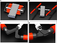
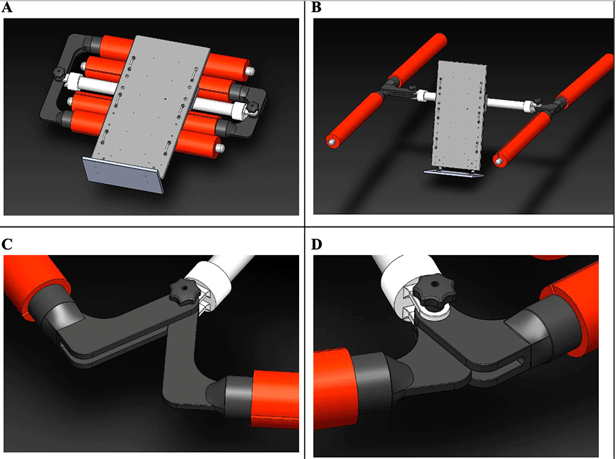
Figure 1: A: The stander in its folded conformation. The board is turned to a horizontal position to allow the buoys to fold underneath. B: The stander in the fully unfolded conformation. The "H-shape" provides even buoyant force distribution. C-D: The L-shaped joints enable the buoys to fold inward without overcomplicating the folding mechanisms of the device.
Patient removal from the device in an emergency must be swift and simple.
The device should be easily portable and compactable so that physical therapists can quickly deploy the device when needed and stow the device when not in use.
Final Approach and Design:
To meet these functional requirements, we created an H-shaped standing frame with four distinct buoys that would elevate the patient in the center. The H-shape is advantageous because it provides an even distribution of buoyant force around the patient. Each buoy consisted of a thin PVC pipe with high density polyethylene foam floats attached. They were attached to the device via custom 3-D printed polymeric pipe joints that were shaped such that the buoys could be easily folded inward to provide easy compactibility. The joints also made it possible for the buoys to be adjusted depending on the space needed by the physical therapist.
In the center of the buoys, we attached a 24'' by 12'' by 0.5'' cushioned ventral support board against which our patient would lay their chest. In order to prevent redness and irritation, the board materials and cushions are made of inert materials that do not react with chemicals found commonly in pools nor the skin of humans. The texture of these materials is smooth, and the cushions are lined with polyvinyl fabric, another inert polymeric material often used for seats and cushions on boats. This board also features webbing guides to secure the patient via cushioned straps, and a foot platform upon which the patient can stand during the physical therapy. The board is attached to the device on a rotational axis, such that the board can be moved from a horizontal to a vertical position during therapy. A locking mechanism is used to secure the board in one of three possible angles in order to increase device foldability. In its most compact form, the device occupied an approximate space of 33'' by 24'' by 8''. When fully unfolded, the device occupied a space of about 45'' by 55'' by 24''.
Outcome:
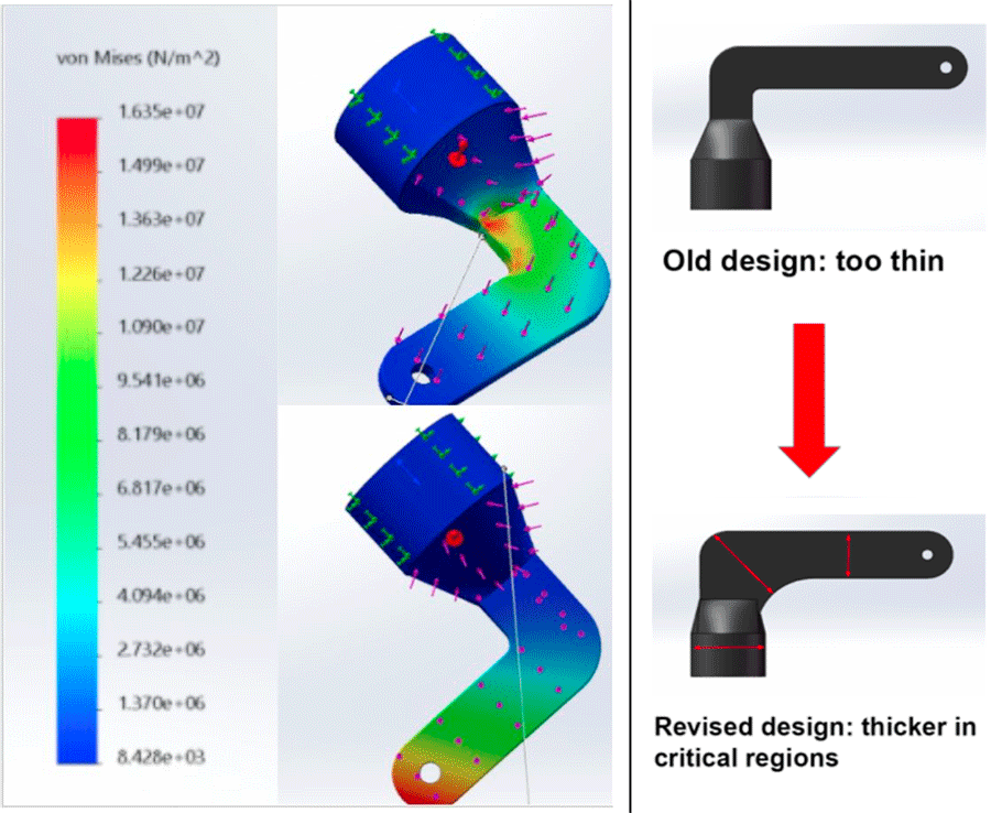
Figure 2: Left: the majority stress is exerted upon the curved portion of the L joint. Colorimetric scales indicate regions of high potential for material failure. Distortion of objects in Solidworks appears drastic, but the software amplifies simulated distortions to an exaggerated level that is unrepresentative of the real part. Right: To address stress accumulation in curved regions of the joint, the next iteration of the joints were made to be thicker in stress critical regions to prevent failure.
Solidworks stress analysis simulations showed that stress accumulates along the curved portion of the custom L joint design and would be indicative of the location of where failure would occur (fig. 2). To address this issue and improve upon the design, the joints were made to be thicker in these critical regions of higher stress accumulation (fig. 2).Furthermore, it is critical to mention the joints are 3D printed from PLA resins and would not be as strong as joints made from custom metal fabrication. Custom metal fabrication wasn't utilized due to costs, but would be heavily recommended for a final product that was to be manufactured.
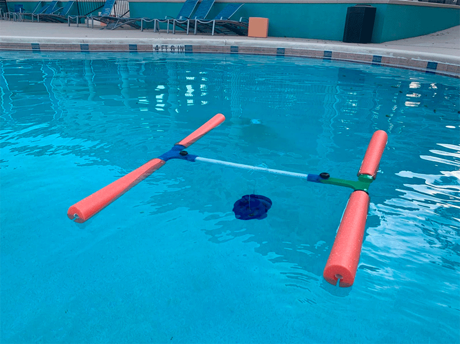
Figure 3: The primary frame and buoys were tested for buoyancy in a swimming pool. The device was able to support a maximum of 36 lbs. Underwater, an average X year old's weight with buoyancy forces under consideration is 13.12 lbs, assuming the position of the X year old is half above water and half underwater due to the position of the board relative to the middle support. Based on the volume of the plates used, each plate provides a net force down of approximately 4 lbs; at 9 weights plates, the device failed, demonstrating that it supports up to 2.6 times the weight of the average X year old.
The current device designed for children from 12-48 months of age who have an average weight range between 21- 27 lbs [23]. Underwater, an average X year old's weight with buoyancy forces under consideration is 13.12 lbs. In-pool buoyancy testing indicated that the current stander prototype supports a maximum weight of 36 pounds when utilizing metallic dumbbells. While we were unable to perform testing with real patients, we believe that the ability of the device to support dense metallic dumbbells in a pool indicates that it should be able to support the weight of children. This device shows promise to be able to support toddlers, but further prototyping and tests are required to confirm this as conclusive.
Cost Analysis:
We monitored our budget throughout the design process. By using the costs of individual components that were used in the final iteration and using industrial average salaries, we were able to estimate the cost of a hypothetical industrial manufacturing process for the production of one HydroStander, including packaging and shipping costs (fig. 4).
There is no guarantee that these cost figures will be entirely accurate after the consideration of a production-level economy of scale. Nonetheless, this cost analysis provides a preliminary assessment on which to base pricing. An industry-standard profit margin of 20 to 30 percent would yield a price range of $227.02 to $245.93 [24]. This pricing places the HydroStander as an extremely price-competitive alternative to several other therapy standers, most of which have prices in the range of thousands of dollars [25].
Figure 4. Cost Analysis For Production of One Stander
The proposed pricing range of $227.02 to $245.93 would allow us to provide a relatively inexpensive physical therapy solution for pediatric patients and physical therapists. The positive effects of this device for enhancing aquatic therapy combined with its highly competitive pricing therefore make this an excellent product for physical therapy clinics to invest in.
Significance:
We are confident that the introduction of this device into the toolkit for physical therapists would have several beneficial effects on the practice of aquatic therapy. Current aquatic therapy methods for children with lower body disabilities require multiple therapists or assistants for safe execution. Our device enables therapists to independently engage with patients without requiring an excessive number of assistants [1], [7], [8], [10], [11]. This can enable clinics to accommodate more patients and perhaps schedule more simultaneous aquatic therapy sessions. We believe that the Aquatin Hydrostander can also enhance the effectiveness of aquatic therapy due to the positive precedent set by the use of standing frames for "dry" physical therapy [13], [15]–[22].
Addendum:
We would like to disclose that the recent COVID-19 pandemic has largely disrupted the progress of this project, and that we were unable to complete a prototype for patient testing for the purposes of the demonstration video. We hope that the content of this design brief has provided enough information to illustrate the potential functionality of our device and the impact that it can have on the field of physical therapy.
References:
J. Prins and D. Cutner, "AQUATIC THERAPY IN THE REHABILITATION OF ATHLETIC INJURIES," Clin. Sports Med., vol. 18, no. 2, pp. 447–461, Apr. 1999, doi: 10.1016/S0278-5919(05)70158-7.
B. E. Becker, "Aquatic therapy: scientific foundations and clinical rehabilitation applications," PM R, vol. 1, no. 9, pp. 859–872, Sep. 2009, doi: 10.1016/j.pmrj.2009.05.017.
P. Tomas-Carus, A. HÄkkinen, N. Gusi, A. Leal, K. Häkkinen, and A. Ortega-Alonso, "Aquatic Training and Detraining on Fitness and Quality of Life in Fibromyalgia," Med. Sci. Sports Exerc., vol. 39, no. 7, pp. 1044–1050, Jul. 2007, doi: 10.1249/01.mss.0b0138059aec4.
A. L. Barker, J. Talevski, R. T. Morello, C. A. Brand, A. E. Rahmann, and D. M. Urquhart, "Effectiveness of Aquatic Exercise for Musculoskeletal Conditions: A Meta-Analysis," Arch. Phys. Med. Rehabil., vol. 95, no. 9, pp. 1776–1786, Sep. 2014, doi: 10.1016/j.apmr.2014.04.005.
T.-J. Wang, B. Belza, F. E. Thompson, J. D. Whitney, and K. Bennett, "Effects of aquatic exercise on flexibility, strength and aerobic fitness in adults with osteoarthritis of the hip or knee," J. Adv. Nurs., vol. 57, no. 2, pp. 141–152, 2007, doi: 10.1111/j.1365-2648.2006.04102.x.
F. B. Wyatt, S. Milam, R. C. Manske, and R. Deere, "The effects of aquatic and traditional exercise programs on persons with knee osteoarthritis," J. Strength Cond. Res., vol. 15, no. 3, pp. 337–340, Aug. 2001.
F. Tripp and K. Krakow, "Effects of an aquatic therapy approach (Halliwick-Therapy) on functional mobility in subacute stroke patients: a randomized controlled trial," Clin. Rehabil., vol. 28, no. 5, pp. 432–439, May 2014, doi: 10.1177/0269215513504942.
H.-G. Cha, Y.-J. Shin, and M.-K. Kim, "Effects of the Bad Ragaz Ring Method on muscle activation of the lower limbs and balance ability in chronic stroke: A randomised controlled trial," Hong Kong Physiother. J., vol. 37, pp. 39–45, Dec. 2017, doi: 10.1016/j.hkpj.2017.02.001.
M. A. Zanon, G. J. Porfírio, R. Riera, and A. L. C. Martimbianco, "Neurodevelopmental treatment approaches for children with cerebral palsy," Cochrane Database Syst. Rev.<, vol. 2015, no. 11, Nov. 2015, doi: 10.1002/14651858.CD011937.
S. C. Chon, D. W. Oh, and J. H. Shim, "Watsu approach for improving spasticity and ambulatory function in hemiparetic patients with stroke," Physiother. Res. Int., vol. 14, no. 2, pp. 128–136, 2009, doi: 10.1002/pri.421.
D. S. V. Hulls, L. K. Walker, and J. M. Powell, "Clinicians' Perceptions of the Benefits of Aquatic Therapy for Young Children with Autism," Phys. Occup. Ther. Pediatr., vol. 26, no. 1–2, pp. 13–22, Jan. 2006, doi: 10.1080/J006v26n01_03.
J. B. Lee, S. B. Kim, K. W. Lee, J. H. Lee, J. G. Park, and S. J. Lee, "Combined Therapy With Functional Electrical Stimulation and Standing Frame in Stroke Patients," Ann. Rehabil. Med., vol. 43, no. 1, pp. 96–105, Feb. 2019, doi: 10.5535/arm.2019.43.1.96.
E. Rivi, M. Filippi, E. Fornasari, M. T. Mascia, A. Ferrari, and S. Costi, "Effectiveness of standing frame on constipation in children with cerebral palsy: a single-subject study," Occup. Ther. Int., vol. 21, no. 3, pp. 115–123, Sep. 2014, doi: 10.1002/oti.1370.
"Effects of a dynamic versus a static prone stander on bone mineral density and behavior in four children with severe cerebral palsy. - Abstract - Europe PMC." https://europepmc.org/article/med/17053680 (accessed Mar. 24, 2020).
K. Taylor, "Factors affecting prescription and implementation of standing-frame programs by school-based physical therapists for children with impaired mobility," Pediatr. Phys. Ther. Off. Publ. Sect. Pediatr. Am. Phys. Ther. Assoc., vol. 21, no. 3, pp. 282–288, 2009, doi: 10.1097/PEP.0b013e3181b175cd.
F. Ferrarello et al., "Passive standing as an adjunct rehabilitation intervention after stroke: a randomized controlled trial," Arch. Physiother., vol. 5, Jul. 2015, doi: 10.1186/s40945-015-0002-0.
A. Logan et al., "Standing Practice In Rehabilitation Early after Stroke (SPIRES): a functional standing frame programme (prolonged standing and repeated sit to stand) to improve function and quality of life and reduce neuromuscular impairment in people with severe sub-acute stroke—a protocol for a feasibility randomised controlled trial," Pilot Feasibility Stud., vol. 4, Mar. 2018, doi: 10.1186/s40814-018-0254-z.
G. Paleg and R. Livingstone, "Systematic review and clinical recommendations for dosage of supported home-based standing programs for adults with stroke, spinal cord injury and other neurological conditions," BMC Musculoskelet. Disord., vol. 16, Nov. 2015, doi: 10.1186/s12891-015-0813-x.
E. Y. Han, J. H. Choi, S.-H. Kim, and S. H. Im, "The effect of weight bearing on bone mineral density and bone growth in children with cerebral palsy," Medicine (Baltimore), vol. 96, no. 10, Mar. 2017, doi: 10.1097/MD.0000000000005896.
[20] S. K. Gibson, J. A. Sprod, and C. A. Maher, "The use of standing frames for contracture management for nonmobile children with cerebral palsy," Int. J. Rehabil. Res. Int. Z. Rehabil. Rev. Int. Rech. Readaptation
J. Goodwin et al., "Understanding frames: A qualitative exploration of standing frame use for young people with cerebral palsy in educational settings," Child Care Health Dev., vol. 45, no. 3, pp. 433–439, 2019, doi: 10.1111/cch.12659.
J. Gillespie, L. Callender, and S. Driver, "Usefulness of a standing frame to improve contraversive pushing in a patient post-stroke in inpatient rehabilitation," Proc. Bayl. Univ. Med. Cent., vol. 32, no. 3, pp. 440–442, May 2019, doi: 10.1080/08998280.2019.1593763.
M. de Onis, WHO child growth standards: length/height-for-age, weight-for-age, weight-for-length, weight-for-height and body mass index-for-age ; methods and development. Geneva: WHO Press, 2006.
The Medicare Payment Advisory Commission, "Chapter 7: An overview of the medical device industry," Report to the Congress: Medicare and the Health Care Delivery System. Jun. 2017.
Adaptive Mall, Standers, Adaptive Mall, Dolgeville, New York, United States, Mar. 25, 2020.
Acknowledgements and References:
We would like to thank Dr. Fuller, Dr. Murfee, and Dr. Rowlinson for their support as faculty instructors as well as their guidance in the completion of this design project. We would also like to thank Mr. Ismael Arroyo for his technical assistance and support that made this project possible. Finally, we would like to thank Lourdes Mengelkoch from UF Health Family Medicine-Magnolia Parke, whose expertise in the field of physical therapy was invaluable for our project. We are funded by the J. Crayton Pruitt Family Department of Biomedical Engineering at the University of Florida and the Herbert Wertheim College of Engineering.